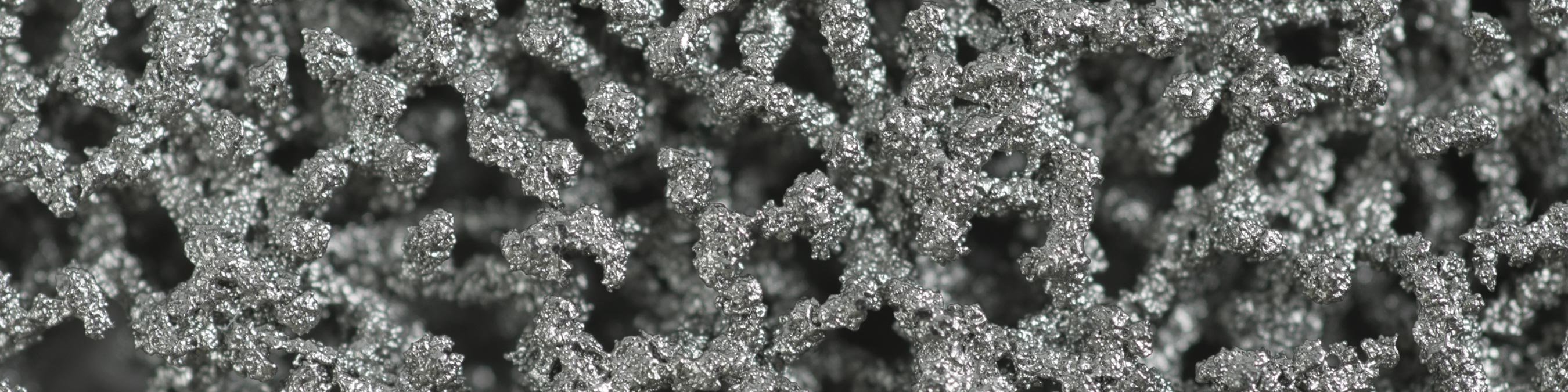
Zimmer Biomet’s OsseoTi Porous Metal Technology uses human CT data in combination with 3D printing technology to build a structure that directly mimics the architecture of human cancellous bone. Using a proprietary manufacturing process, constructs are printed layer by layer to build a fully-integrated component with solid and porous regions. This process maintains the consistent porosity and strength necessary to facilitate tissue in-growth and implant stability.1*
OsseoTi® Porous Metal Technology


OsseoTi Porous Metal Technology features:
- A porous architecture that has demonstrated excellent integration with host bone as early as 4 weeks in an animal study1*
- Architecture directly mimics the structure of human cancellous bone
- Porosity of approximately 70%
- Average pore size of 475 microns, which facilitates cell migration, vascularization, and tissue ingrowth1*
- Material strength between that of cancellous and cortical bone, which facilitates biologic fixation and loading of surrounding bone 1,2*
*Animal studies are not necessarily indicative of clinical performance
Zimmer Biomet’s OsseoTi® Porous Metal Technology is made from the highly biocompatible Ti6A14V alloy material that has excellent corrosion resistance and a proven history of clinical success.3,4 Mimicking the porous structure of human cancellous bone, the physical properties of OsseoTi Porous Metal are designed for the potential of bone integration.5 This porous metal structure facilitates tissue ingrowth and remodeling, vascularization, and very close attachment between bone and the metal. Zimmer Biomet’s OsseoTi Porous Metal Technology has the potential to facilitate bone integration for orthopedic devices.
* Animal studies are not necessarily indicative of clinical performance
- Data on file at Biomet. Bench test results not necessarily indicative of clinical performance. Laboratory testing.
- Gupta, G. OsseoTi Porous Metal For Enhanced Bone Integration: an Animal Study. Biomet Form No. BMET0718.0-GBL. Animal studies are not necessarily indicative of clinical performance.
- Long M, Rack HJ. Titanium alloys in total joint replacement – A materials science perspective. (1998) Biomaterials 19-1621-1639.
- Woodell-May J, Kumar M. In vitro comparison of cell proliferation on Ti6A14V and Tantalum Metal. ORS2007. Poster # 1578.
- Data on file at Biomet, Inc. Protocol #12-04/ 12-07.
Legal Manufacturer:
Biomet Orthopedics
56 East Bell Drive
P.O. Box 587
Warsaw, Indiana 46581 USA