Welcome to Zimmer Biomet Latin America
Welcome to Zimmer Biomet Latin America, where our commitment to advancing healthcare is built on a legacy of innovation and success. Guided by our mission to alleviate pain and improve quality of life, we are dedicated to providing world-class solutions that empower patients, support healthcare professionals, and foster positive outcomes across the region. Through our advanced technologies and passionate team, we strive to make a meaningful difference in the lives of people throughout Latin America.
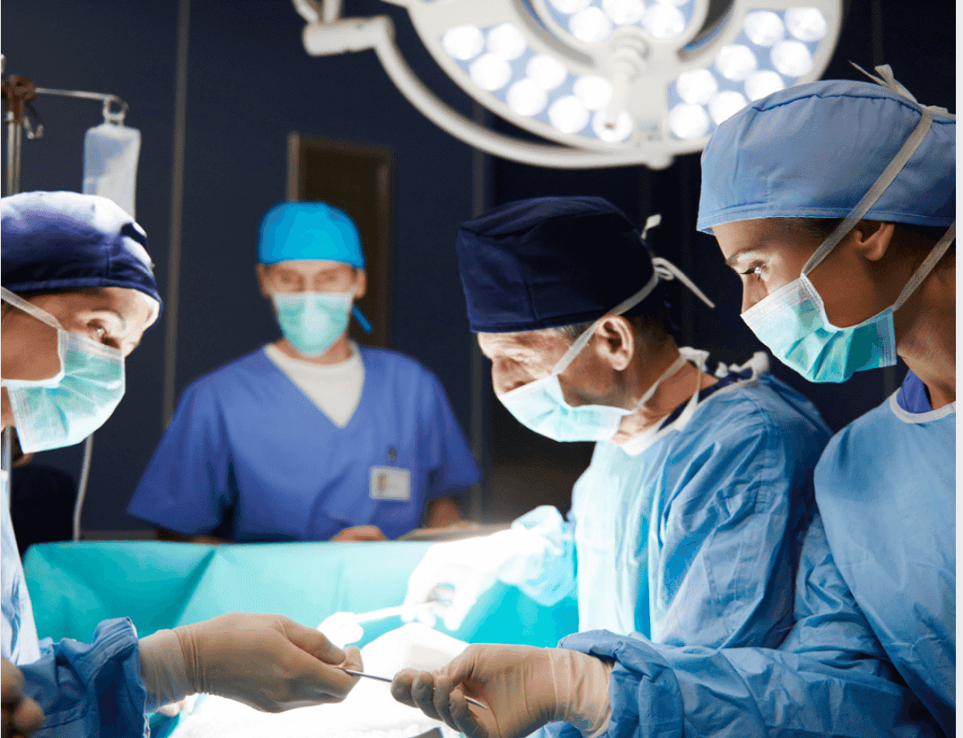
For Medical Professionals
As a global medical technology leader, we push the boundaries of innovation to redefine what's possible across all stages of the patient journey.
For Patients & Caregivers
At Zimmer Biomet, our innovations help treat patients suffering from a variety of disorders and injuries involving our bones and joints. And, while we love and believe in our amazing products, we get up every day and do what we do so that you become you again.
Contact Zimmer Biomet Latin America
All content herein is protected by copyright, trademarks and other intellectual property rights, as applicable, owned by or licensed to Zimmer Biomet or its affiliates unless otherwise indicated, and must not be redistributed, duplicated or disclosed, in whole or in part, without the express written consent of Zimmer Biomet.